|
The open cavity presents the advanced stage of the carious lesion. Usually the disease process is active, and is progressing at an accelerated rate. This results from the larger receptacle for retention of food material which undergoes decomposition within the cavity. If fermentable carbohydrates are present acids are produced. Such acids decalcify tooth substance - both dentin and enamel - and thus promote advancement of the lesion.
The bottom and sidewalls of the cavity are covered with a pad or film of bacterial material consisting largely of filamentous types of organisms with one oend attached to the decaying dentin, the other extending outward towards the surface of the pad. At the surface there are the growing ends and fruiting heads of the long rods and filaments composing the pad, and also a great variety or other organisms from the decomposing food material within the cavity.
The purpose of this paper is to report the frequency of the presence or Leptothrix racemosa, based upon finding the fruiting heads of this organism, in cavities in a miscellaneous collection of extracted tooth specimens.
Materials and Methods
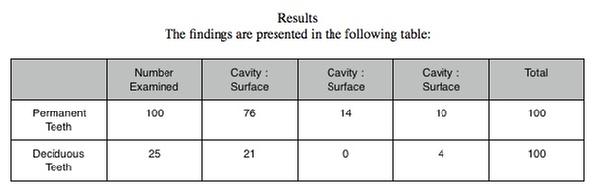
One hundred permanent teeth (75 molars, 16 bicuspids, 4 cuspids, 5 upper centrals) with large open cavities were selected from a miscellaneous collection of extracted teeth received from extraction clinics in New Orleans, and preserved in 10% formalin. They were rinsed in water, stained in crystal violet (0.5% in water) for about one minute or longer and then rinsed again in water. Under the dissecting microscope. with appropriate lighting from above, material was picked from the bottom or sidewalls of the cavity, teased out somewhat in a droplet of 50% glycerine (in water) on a slide and covered with a one-fourth size cover glass. The preparation is now ready for microscopic examination.
The method of handling stained tooth specimens and the facilities previously described for another purpose are also quite satisfactory for this purpose. Especially useful for removing material from the tooth for microscopic preparations in studying the composition of the film pad, is a teasing needle made by grinding to spade or spatula shape a No. 10 silver steel sewing needle driven eye end into the four inch length of three-sixteenths inch wooden doweling. This instrument is especially useful to run under and lift (dig) off bacterial film without breaking it up too much, as occurs when it is scraped off with other instruments.
Similarly made teasing needles, ground to very sharp, keen points, are best for teasing apart under the dissecting microscope, as required, lumps of the film pack, to search for the fruiting heads of L. racemosa.
Prepared in this way many of the small particles of filamentous material chance to be mounted so the part from the outer surface of the pad where the fruiting heads are found lies in one direction and the deeper part, which was attached to the tooth, lies in the opposite direction. Many of the rods and filaments lie more or less lengthwise in the preparation. Usually it is not difficult to know which part of a particle represents the outer surface of the pad, when it was in situ. The fact that the pad is composed of filamentous type organisms is readily observed. If the specimen is stained for only a short period (less than one minute) the filaments and fruiting heads at the surface are heavily stained, whereas the filaments and stems are stained less or not at all, in the deeper part.
Twenty-five deciduous molars were selected from the same miscellaneous collection and were examined in the same way for fruiting heads of L. racemosa.
Whenever the first preparation from a given specimen in either lot was found negative or unsuitable a second and sometimes a third was made and examined.
At the time preparations were made and examined from cavities, preparations were also made and examined from heavy bacterial film on the surface of the tooth. With experience one soon learns to pick material from the surface (depressions and protected locations) where fruiting heads are most abundant and are most likely to be found.
|